fundamentals
What is Video Laryngeal Stroboscopy?
Laryngeal stroboscopy is a specialized form of video endoscopy with its main function to visualize vocal fold vibration as a means of evaluating voice function. Most often performed by an otolaryngologist or speech-language pathologist, this technique allows for diagnosis and management of a variety of conditions related to abnormalities in vibratory function.
Principles of Stroboscopy
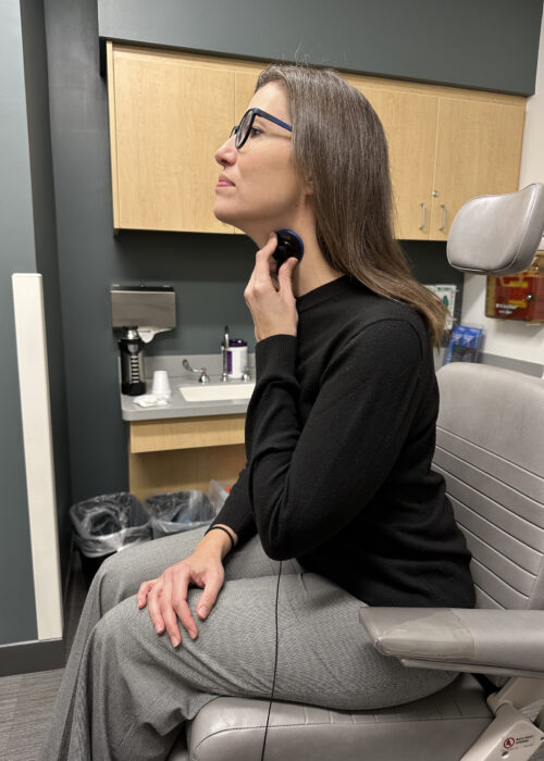
Stroboscopic light uses the principle of the retention of light on the human retina. A single image will be retained on the retina for 200 milliseconds and when a series of individual images are presented in sequence to the eye, the brain will fuse them, leading to the perception of motion (Talbot’s Law). Modern stroboscopy machines are able to detect the fundamental frequency of vibration of a subject’s voice (via a contact microphone placed on the neck) and then turn the strobing light frequency several hertz (1-3) above it thus leading to the appearance of slow pseudo-motion where individual frames of the glottic cycle are perceived to be fused together in apparent motion.
Initially, rigid endoscopes with bulky cameras were used to perform laryngeal stroboscopy. Later developments in flexible fiberoptic endoscopy led to the ability to combine flexible technology with a strobe light. Exams could now be conducted transnasally with a patient’s connected speech. Although the fiberoptic technology provided ease of examination, there was a notable decline in image quality.
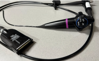
In the modern era of stroboscopy, charged coupled device (CCD) cameras are mounted onto the tip of endoscopes offering greater image resolution. This technology, initially used on the tips of gastroscopes, has been adopted for our indirect laryngoscopes by manufacturers such as Olympus and Pentax and has gained widespread use in the voice professional community.
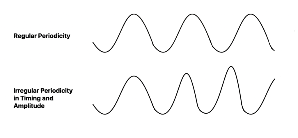
Despite its clinical contributions, stroboscopy has some disadvantages. If the patient’s vocal folds vibrate in a non-quasiperiodic manner (such as in patients with intermittent vibratory abnormalities) stroboscopy can fail to adequately evaluate the patient. Consequently, high speed video imaging was developed for such scenarios. This equipment however can be cost prohibitive and currently remains largely utilized in the research arena
Equipment and Clinical Operation
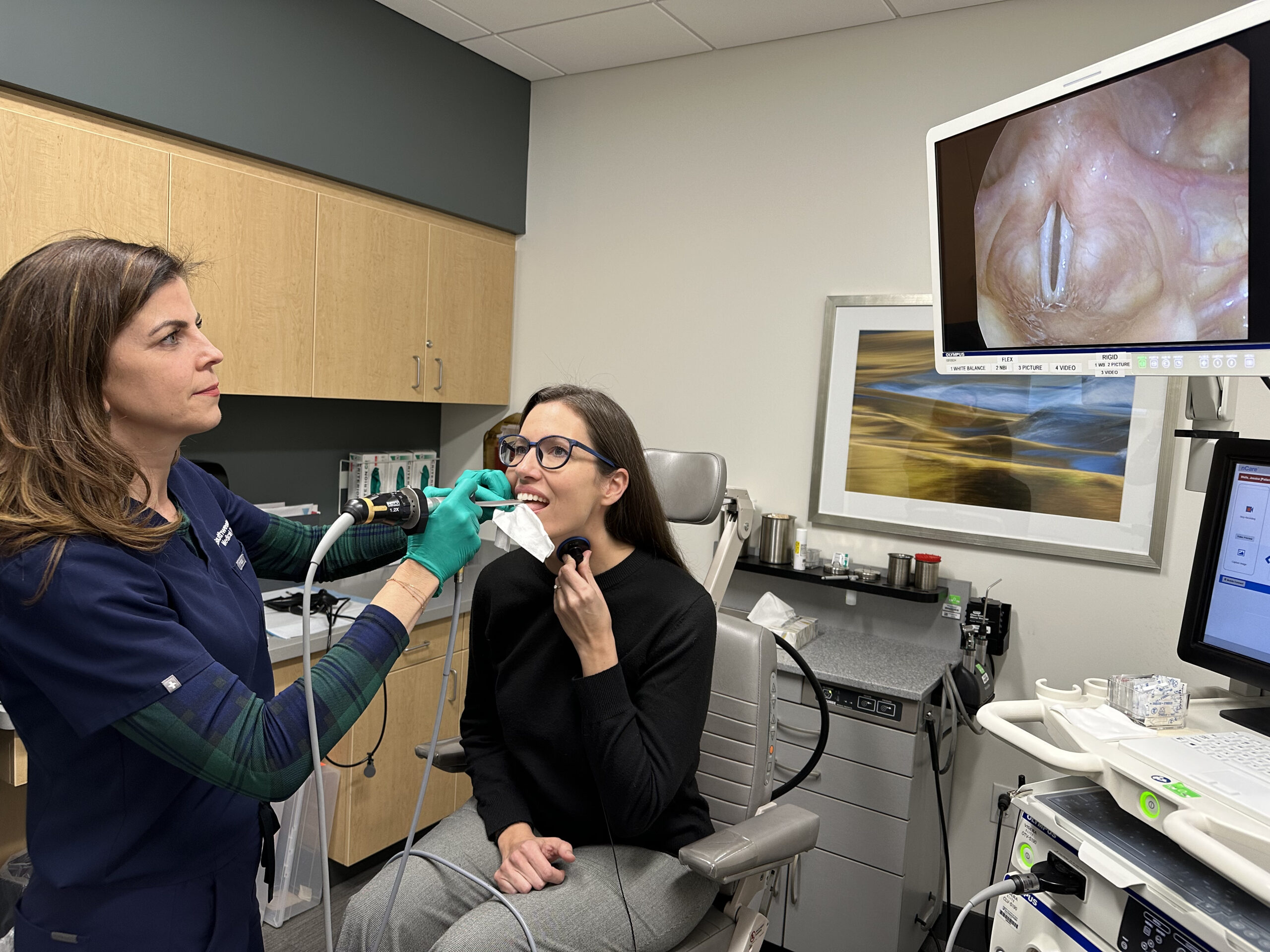
The main aim of this website is in teaching laryngeal stroboscopy analysis, however, below is a brief introduction to the typical set up of a clinical stroboscopy room.
Examination Chair
A wide chair with a foot rest, moveable headrest and the ability to raise and lower the height will facilitate patient and provider comfort during examination.
Topical Anesthetic
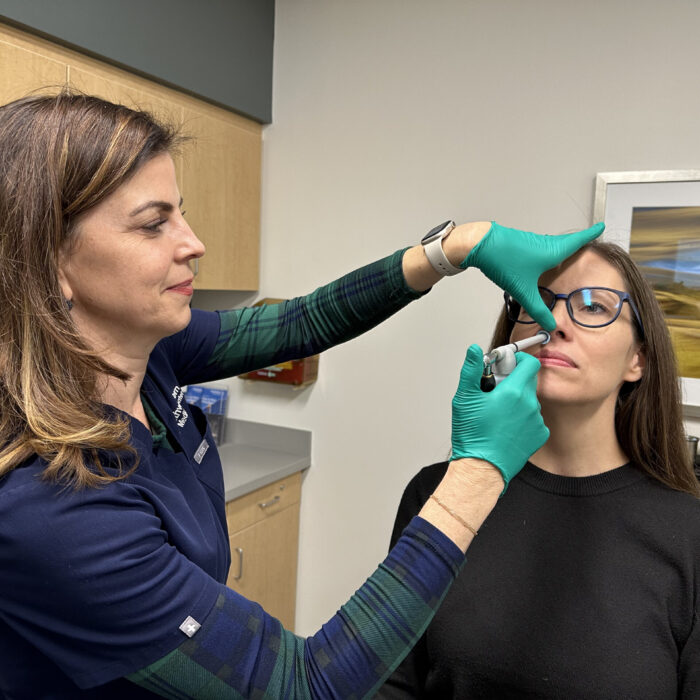
Nasal decongestant spray and topical anesthesia can be used to improve patient comfort during stroboscopy. Topical anesthesia can be applied to the nasal cavity, nasopharynx, and oropharynx. Nasal decongestants are applied to the nasal cavity. After application, wait for adequate anesthesia/vasoconstriction to take effect before proceeding with flexible laryngoscopy. Medication selection for laryngeal anesthesia (lidocaine, pontocaine, cetacaine) or decongestant (neosynephrine, oxymetazoline) is up to the endoscopist. Excessive use of topical anesthesia can lead to increased secretions and can cause systemic toxicity.
Patient and Stroboscopist Positioning
The patient should be in an upright forward position, with the neck slightly flexed and head slightly extended with feet firmly placed flat on the footrest. The examination chair should be raised or depressed based on the height of the examiner to avoid strain and fatigue.
Stroboscope light source
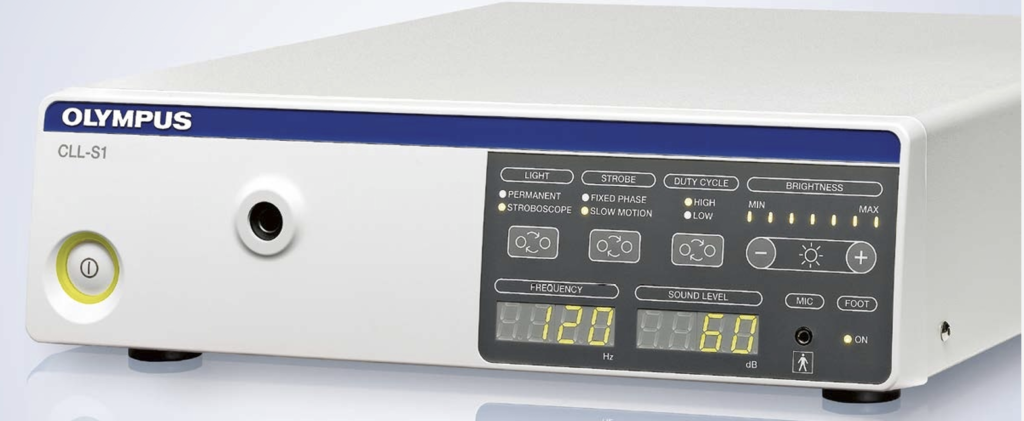
this provides the light output. Many modern machines have a combination of halogen and xenon light for non-strobe and strobe type examinations. Sound is then transmitted from the patient to the stroboscope via a microphone or contact stethoscope. Circuitry within the strobe machine then derives the fundamental frequency from the voice. A foot pedal is helpful for the endoscopist to control various aspects of the strobe light and trigger a digital recording when ready.
Recording device and Video reviewing software
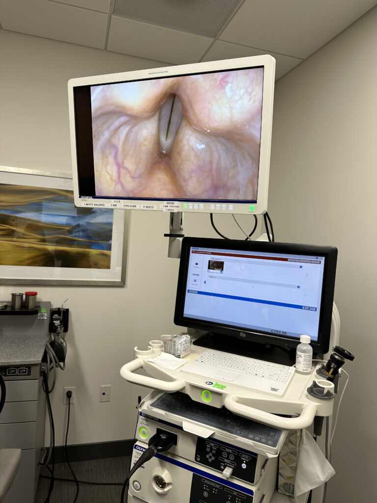
CCD camera technology on distal tip chip scopes provide excellent image quality. The ability to record and easily access stroboscopy videos is paramount and can be saved either directly on a local processing hard drive or uploaded to a local server or cloud technology.
Laryngoscope
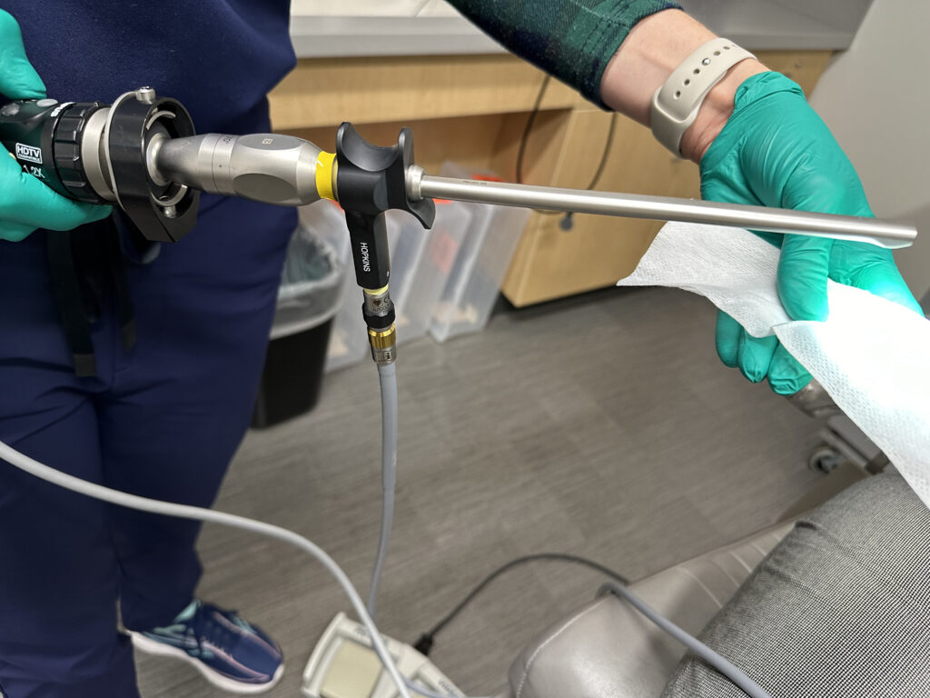
Various types of flexible laryngoscopes can be used for laryngeal stroboscopy, however in recent years, distal tip chip scopes have replaced fiberoptic technology. Many clinicians prefer rigid telescopes with angled tips (70 or 90 degree) for transoral visualization which can provide sharper image quality and greater magnification of the laryngeal structures.
Output monitor
Many offices have several high-definition output monitors, one for the endoscopist to view during the exam and one for the patient to view directly in front of the patient. The important factor is that the exam is properly recorded digitally and able to be reviewed with the patient, including frame by frame analysis.

